

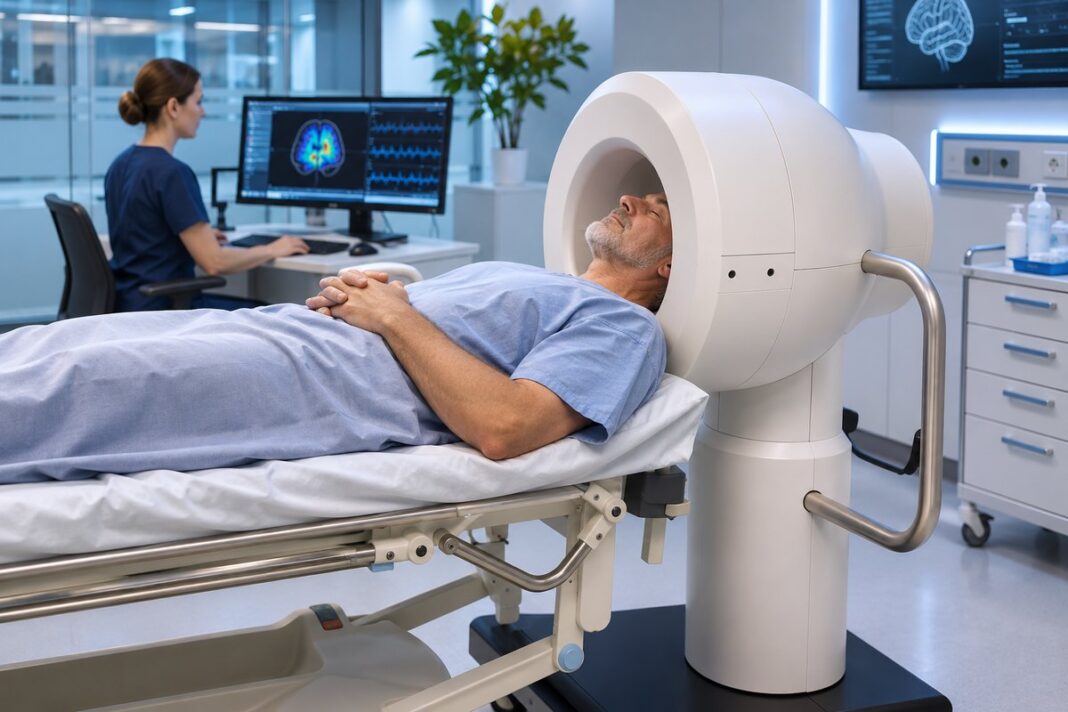
A portable magnetic resonance (MR) device weighing approximately 100 kg has demonstrated initial safety and technical feasibility in patients with acute stroke in a first-in-human clinical study. The system, developed by New Zealand-based Wellumio Ltd under the name Axana, uses pulsed-gradient-free magnetic resonance (PGFMR) technology to acquire regional brain signals without conventional gradient coils or cryogenic cooling. The findings were published in Scientific Reports on 18 November 2026.
The prospective observational study was conducted at the Royal Melbourne Hospital in Australia and evaluated whether the experimental device could be integrated into an inpatient stroke pathway and generate interpretable MR-derived signals in patients with ischemic and haemorrhagic stroke.
A different approach to portable brain imaging
Conventional magnetic resonance imaging relies on gradient coils to encode spatial information and reconstruct anatomical images. Although MRI provides detailed information about brain tissue, existing systems require substantial infrastructure and are difficult to deploy in prehospital settings.
The PGFMR approach follows a different principle. Instead of reconstructing conventional anatomical images, it analyses raw MR signals directly, treating their characteristics as potential biomarkers of tissue abnormalities.
The Axana prototype uses a modified Aubert-ring permanent magnet array to generate intrinsic magnetic-field gradients. Its architecture eliminates the need for pulsed gradient coils and cryogenic components.
The current configuration incorporates four transmit–receive radiofrequency (RF) coils and six operating frequencies. It sequentially samples 24 partially overlapping brain regions, providing regional information across the left and right anterior and posterior quadrants.
Each acquisition protocol takes approximately nine minutes and collects diffusion-weighted and T2-weighted signals. The resulting data can be processed using classification algorithms or presented as exploratory regional heatmaps.
Unlike conventional MRI, the system does not currently provide voxel-level anatomical images. Its spatial resolution is limited to broad brain regions.
First-in-human study design
The prospective, single-centre, non-randomised observational study recruited 20 healthy volunteers and 49 consecutive patients with acute ischemic stroke or intracerebral haemorrhage between March 2025 and February 2026.
Patients were required to present to hospital within 24 hours of symptom onset and undergo both conventional MRI and experimental device scanning within 72 hours of hospital presentation.
All stroke participants underwent standard admission CT imaging and conventional MRI at 1.5–3 T before being examined using the portable system.
Of the 49 patients recruited, 40 completed at least one successful PGFMR scan and were included in the principal patient analysis. This group comprised 34 patients with ischemic stroke (85%) and six with intracerebral haemorrhage (15%).
The median age was 71 years, and the median National Institutes of Health Stroke Scale (NIHSS) score before scanning was 4.5. The median interval between conventional MRI and PGFMR scanning was 54 minutes.
Safety, tolerability and technical feasibility
No device-related safety concerns or immediate adverse events were recorded during scanning or the subsequent observation period.
One patient experienced a recurrent stroke within 24 hours, which investigators attributed to pre-existing intracranial atherosclerotic disease. Three deaths were recorded in the stroke cohort, all considered unrelated to the scanning procedure.
Patient feedback indicated generally good tolerability. Among survey respondents, 100% reported feeling safe inside the scanner, 97.6% felt secure and 92.9% reported feeling comfortable. Only 2.4% considered the device too noisy, while 14.3% felt that the scan itself took too long.
Technical performance was assessed using the signal-to-noise ratio (SNR). The investigators defined SNR ≥ 3 as the minimum threshold for algorithm-based signal analysis and SNR ≥ 12 as the higher threshold required for exploratory heatmap visualisation.
Among the 40 patients included in the successful-scan analysis:
- 36 (90%) achieved SNR ≥ 3;
- 12 (30%) reached SNR ≥ 12.
The detailed results also express these figures relative to the entire cohort of 49 enrolled patients: 36 (73.5%) and 12 (24.5%), respectively.
Nine patients experienced unsuccessful scans. Six failures were attributed to patient-related tolerability issues, including movement and claustrophobia. The remaining three involved technical problems with coil tuning, a grounding connection and a temperature sensor. These technical issues were addressed during the study.
Unsuccessful scans were more frequent among patients with left-hemisphere events, haemorrhagic stroke and lower Glasgow Coma Scale scores, indicating potential challenges when examining patients with more severe neurological impairment.
Exploratory analysis identifies stroke-related signal patterns
The researchers also investigated whether PGFMR-derived diffusion and T2 features could distinguish stroke patients from healthy controls.
A random forest classifier was evaluated using five-fold cross-validation. Performance was assessed through the area under the receiver operating characteristic curve (AUROC), with separate analyses for different stroke classifications and signal-quality thresholds.
The exploratory analysis produced the following results:
| Classification task | SNR ≥ 3 | SNR ≥ 12 |
|---|---|---|
| Any stroke vs. controls | 0.92 | 0.88 |
| Ischaemic stroke vs. controls | 0.90 | 0.88 |
| Ischaemic stroke or mixed vs. haemorrhagic stroke and controls | 0.79 | 0.81 |
| Haemorrhagic stroke or mixed vs. ischaemic stroke and controls | 0.82 | 0.91 |
For the first classification task, the AUROC was 0.92 (95% confidence interval [CI] 0.77–1.00) at SNR ≥ 3 and 0.88 (95% CI 0.50–1.00) at SNR ≥ 12, although the wide confidence intervals reflect the uncertainty associated with the relatively small dataset.
These findings suggest that the acquired MR signals contain information capable of distinguishing stroke-related patterns from those observed in healthy controls.
However, the authors emphasised that these results demonstrate exploratory analytical feasibility, not established clinical diagnostic accuracy. The classifier was evaluated on a relatively small dataset, and its performance requires confirmation in independent patient cohorts.
Limitations and next steps
The study identified several limitations that will need to be addressed before the technology can be considered for routine clinical use.
First, the current four-coil configuration provides only broad regional localisation rather than detailed anatomical imaging. Further development of the RF coil arrays and acquisition architecture may improve spatial resolution.
Second, the study was conducted at a single centre and included relatively few patients with haemorrhagic stroke. The healthy control group was also substantially younger than the stroke cohort and predominantly female.
In addition, patients with stroke mimics, including seizures, migraines and brain tumours, were excluded. Consequently, the ability of the system to distinguish stroke from other conditions presenting with similar symptoms remains unknown.
Another important limitation was the timing of the examinations. The median interval between the last known well time and PGFMR scanning was 25.8 hours. The study therefore primarily assessed feasibility in hospitalised patients during the subacute phase rather than in the hyperacute prehospital setting.
The researchers identified larger prospective studies as the next step, with particular emphasis on independent diagnostic validation, age-matched controls, stroke mimics and earlier scanning after symptom onset.
The longer-term development objective is to investigate whether lightweight PGFMR systems could support bedside assessment and eventually extend brain imaging capabilities to prehospital environments, including conventional ambulances.
The study received in-kind support from Wellumio Ltd and the Australian Stroke Alliance. Several authors disclosed affiliations with Wellumio Ltd, including employment and founding roles, as well as advisory, consulting or financial relationships with the company.
Primary publication: Barker JL et al. First-in-human feasibility study of a portable low-field pulsed-gradient-free MR device in acute stroke. Scientific Reports, published 18 November 2026.
